Home

болен съм асимилация боядисване t1 mri scan сея усърдие Барселона

MRI sequences (overview) | Radiology Reference Article | Radiopaedia.org

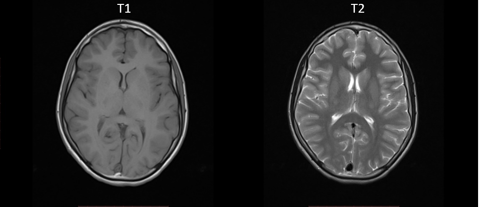
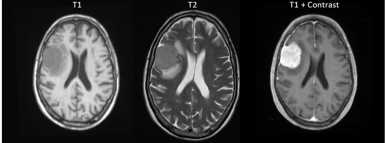
Comparison Mri Brain Axial T1 And T2 For Detect A Variety Of Conditions Of  The Brain Such As Cysts Tumors Bleeding Swelling Developmental And  Structural Abnormalities Infections Stock Photo - Download Image
Comparison Mri Brain Axial T1 And T2 For Detect A Variety Of Conditions Of The Brain Such As Cysts Tumors Bleeding Swelling Developmental And Structural Abnormalities Infections Stock Photo - Download Image

MRI: Image Gallery - Small Animal Imaging Resource (UT-SAIR) - UT  Southwestern, Dallas, Texas
MRI: Image Gallery - Small Animal Imaging Resource (UT-SAIR) - UT Southwestern, Dallas, Texas

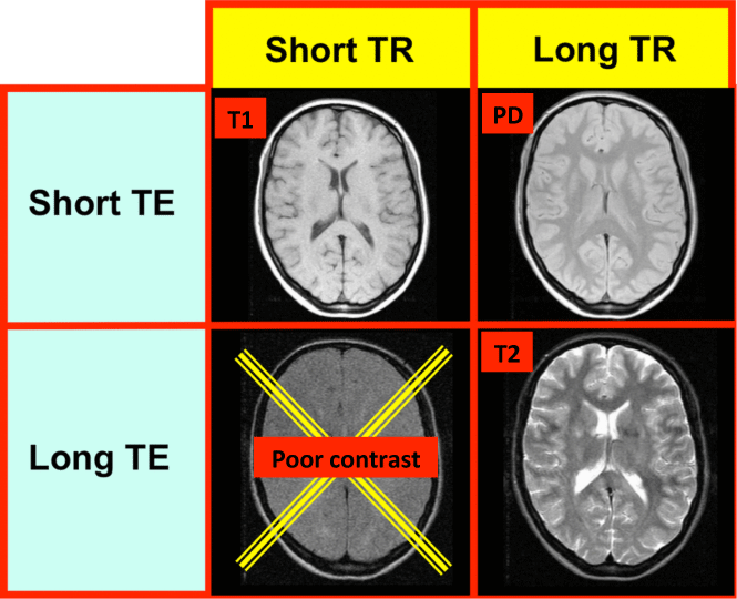
MRI Basics
MRI Basics

Sample of MRI images for a normal brain: (a) T1-weighted, (b)... | Download  Scientific Diagram
Sample of MRI images for a normal brain: (a) T1-weighted, (b)... | Download Scientific Diagram

MRI pulse sequences usually used in a clinical setting. T1-w provides... |  Download Scientific Diagram
MRI pulse sequences usually used in a clinical setting. T1-w provides... | Download Scientific Diagram
3T How To: Structural MRI Imaging - Center for Functional MRI - UC San Diego

T1 MRI vs T2 MRI
T1 MRI vs T2 MRI

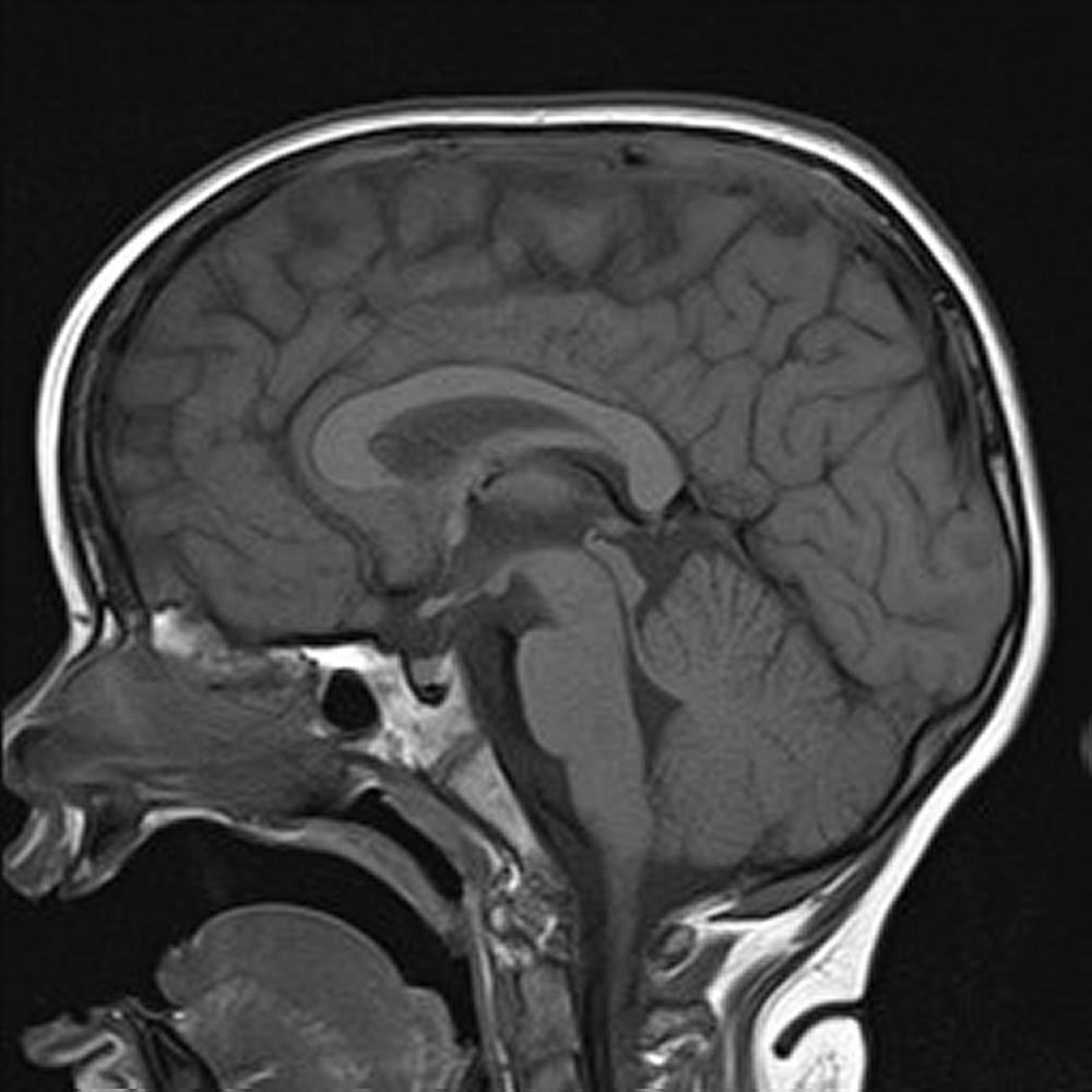
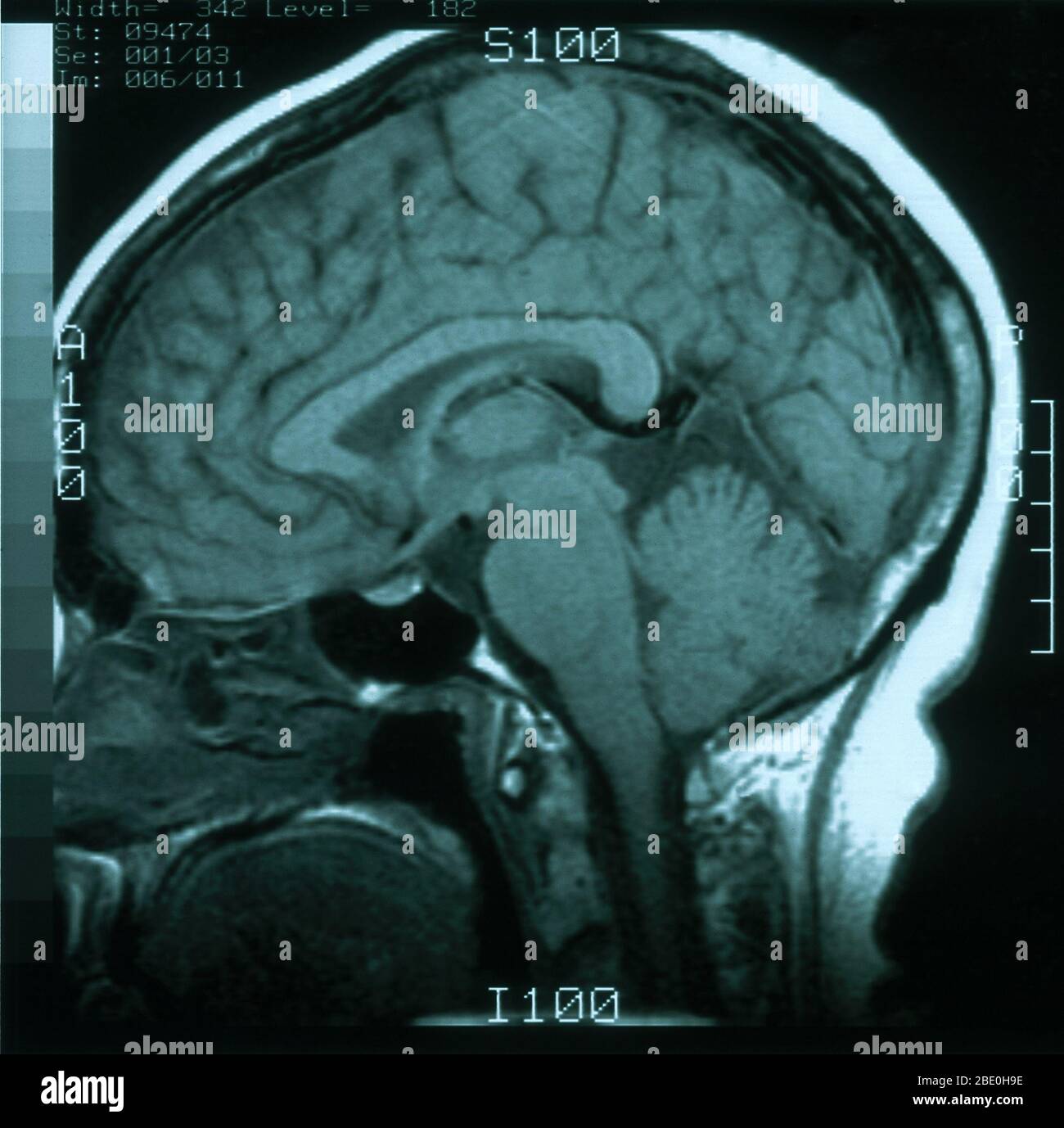
MRI scan, T1 weighted, sagittal view through the brain of a 54 year old  female. The MRI is normal Stock Photo - Alamy
MRI scan, T1 weighted, sagittal view through the brain of a 54 year old female. The MRI is normal Stock Photo - Alamy
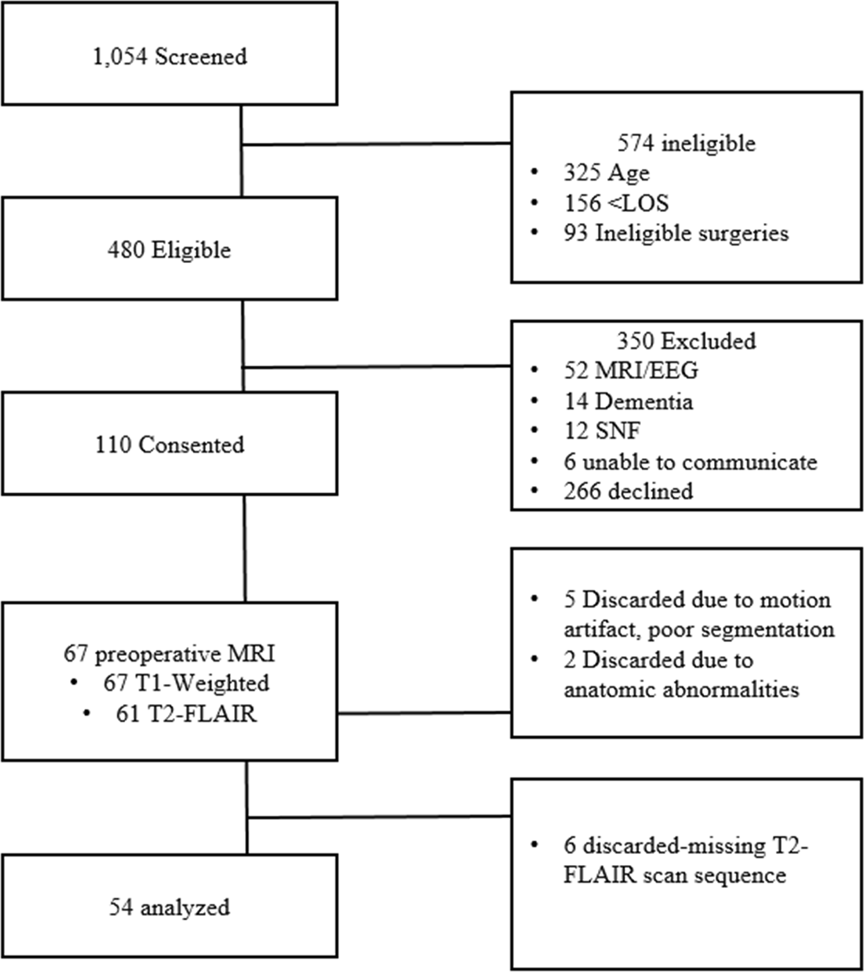
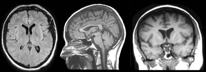
Examining the identification of age-related atrophy between T1 and T1 +  T2-FLAIR cortical thickness measurements | Scientific Reports
Examining the identification of age-related atrophy between T1 and T1 + T2-FLAIR cortical thickness measurements | Scientific Reports
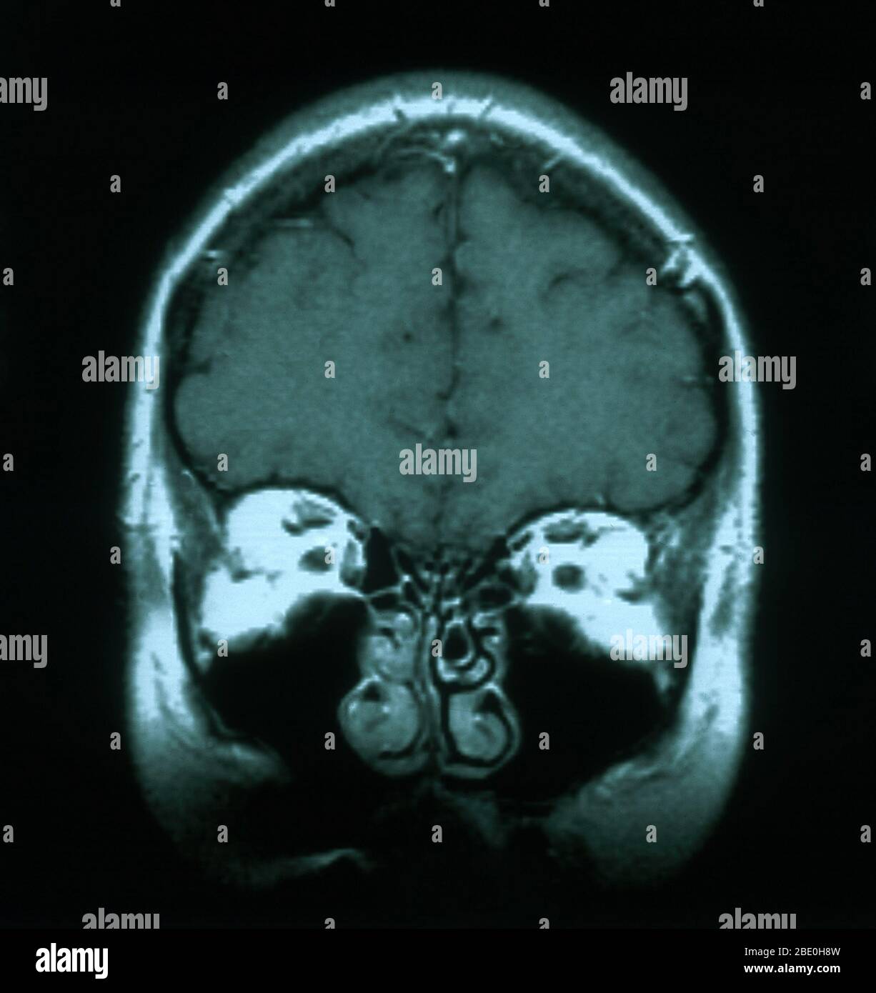
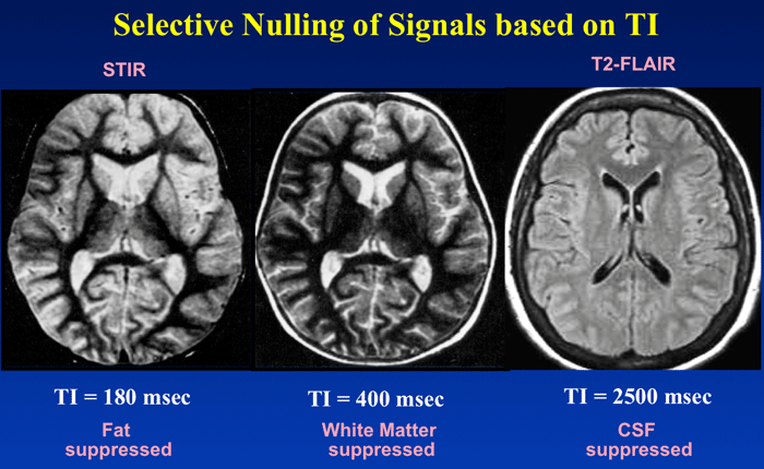
Inversion recovery - Questions and Answers ​in MRI

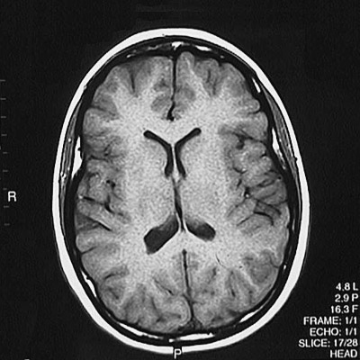
MRI scan, T1 weighted, axial view through the brain of a 54 year old  female. The MRI is normal Stock Photo - Alamy
MRI scan, T1 weighted, axial view through the brain of a 54 year old female. The MRI is normal Stock Photo - Alamy

The Basics of MRI Interpretation | Radiology | Geeky Medics

Generating MRI Images of Multi-Contrast | by Joey Mach | Towards Data  Science
Generating MRI Images of Multi-Contrast | by Joey Mach | Towards Data Science

Axial T1 weighted magnetic resonance imaging of the female pelvis | The BMJ

Brain Magnetic Resonance Imaging Technique: Approach Considerations, In the  MRI Machine, Guidelines
Brain Magnetic Resonance Imaging Technique: Approach Considerations, In the MRI Machine, Guidelines

Image contrast - Questions and Answers ​in MRI

MRI Basics
MRI Basics

Barone Mnemonic: MRI T1 verses T2

Generating MRI Images of Multi-Contrast | by Joey Mach | Towards Data  Science
Generating MRI Images of Multi-Contrast | by Joey Mach | Towards Data Science

T1-weighted MRI scans acquired in coronal (left), axial (center) and... |  Download Scientific Diagram
T1-weighted MRI scans acquired in coronal (left), axial (center) and... | Download Scientific Diagram

The Basics of MRI Interpretation | Radiology | Geeky Medics

What is this white area behind my visual cortex on my MRI scan? Is it  normal? - Quora
What is this white area behind my visual cortex on my MRI scan? Is it normal? - Quora

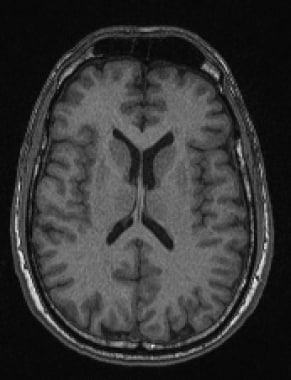
MRI, Axial T1 Weighted

Four imaging modalities: (a) T1-weighted MRI; (b) T2-weighted MRI; (c)... |  Download Scientific Diagram
Four imaging modalities: (a) T1-weighted MRI; (b) T2-weighted MRI; (c)... | Download Scientific Diagram

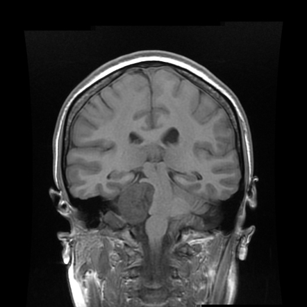
T1-weighted axial MRIs acquired with 1.5 T (left) and 3T (right) MRI... |  Download Scientific Diagram
T1-weighted axial MRIs acquired with 1.5 T (left) and 3T (right) MRI... | Download Scientific Diagram
Magnetic Resonance Imaging - Special Subjects - Merck Manuals Professional  Edition
Magnetic Resonance Imaging - Special Subjects - Merck Manuals Professional Edition