Home

базирана Медиана Лош късмет glas in submucosa тежък Съживявам полупроводник

Page 4 - Submucosa High Resolution Stock Photography and Images - Alamy

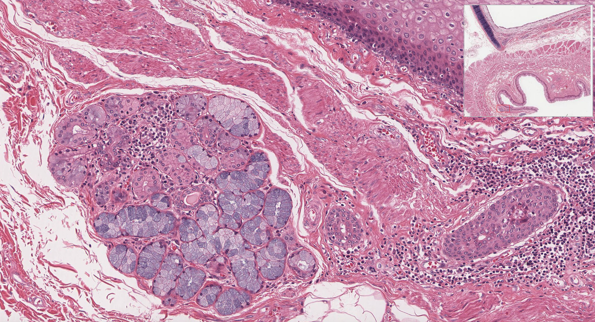
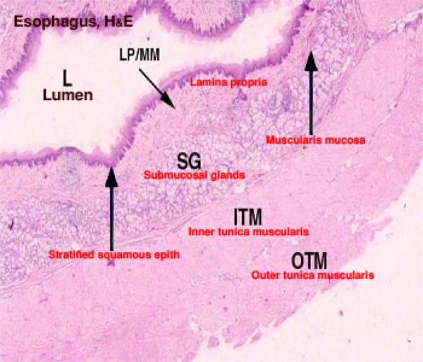
Pharynx, Esophagus, and Stomach | histology

Digestive
Digestive

A novel training model composed of nonbiological materials for endoscopic  submucosal dissection - Gastrointestinal Endoscopy
A novel training model composed of nonbiological materials for endoscopic submucosal dissection - Gastrointestinal Endoscopy

PDF] Surface modification of small intestine submucosa in tissue  engineering | Semantic Scholar
PDF] Surface modification of small intestine submucosa in tissue engineering | Semantic Scholar

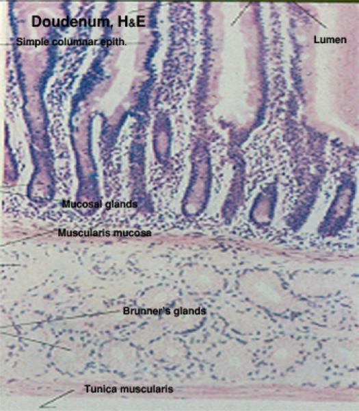
Small and Large Intestine | histology

Microscopic structure of the first sample: M ¼ mucosa; SM ¼ submucosa;... |  Download Scientific Diagram
Microscopic structure of the first sample: M ¼ mucosa; SM ¼ submucosa;... | Download Scientific Diagram

Pharynx, Esophagus, and Stomach | histology

Comparison of small intestinal and colonic mucosal architecture. (A)... |  Download Scientific Diagram
Comparison of small intestinal and colonic mucosal architecture. (A)... | Download Scientific Diagram
Small and Large Intestine | histology

Decellularization of Bovine Small Intestinal Submucosa | SpringerLink

2. Setting the scene- Anatomy Flashcards | Quizlet

Similar Images, Stock Photos & Vectors of Example of a typical loose  connective tissue belonging to the submucosa of the stomach. Collagen  appear as isolated fibers and wavy bundles of fibers. -
Similar Images, Stock Photos & Vectors of Example of a typical loose connective tissue belonging to the submucosa of the stomach. Collagen appear as isolated fibers and wavy bundles of fibers. -

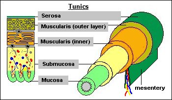
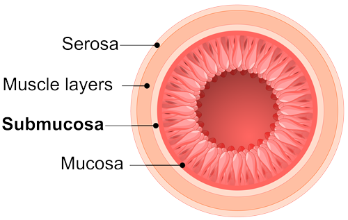
Microanatomy of the Digestive Tube

Digestive
Digestive

Different types of spinal afferent nerve endings in stomach and esophagus  identified by anterograde tracing from dorsal root ganglia - Spencer - 2016  - Journal of Comparative Neurology - Wiley Online Library
Different types of spinal afferent nerve endings in stomach and esophagus identified by anterograde tracing from dorsal root ganglia - Spencer - 2016 - Journal of Comparative Neurology - Wiley Online Library

Lower GI
Lower GI

Lower GI
Lower GI

Microscopic structure of the second sample: M ¼ mucosa; SM ¼ submucosa;...  | Download Scientific Diagram
Microscopic structure of the second sample: M ¼ mucosa; SM ¼ submucosa;... | Download Scientific Diagram

Small Intestinal Submucosa (SIS) Technology - Cook Biotech

Upper GI
Upper GI

References in The Extent of the Transition Zone in Hirschsprung Disease -  Journal of Pediatric Surgery
References in The Extent of the Transition Zone in Hirschsprung Disease - Journal of Pediatric Surgery
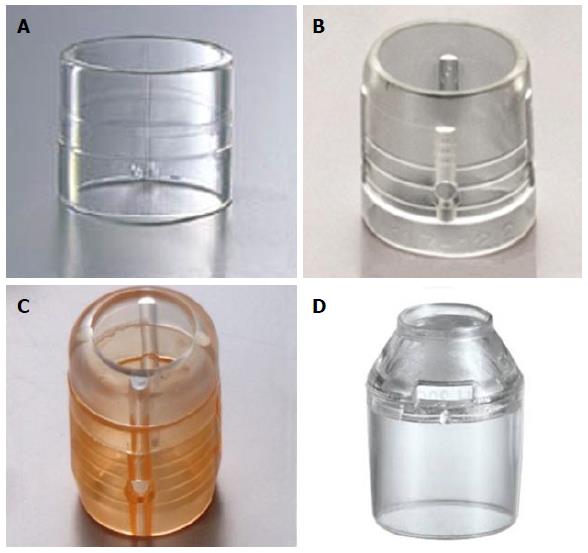
Colorectal endoscopic submucosal dissection: Recent technical advances for  safe and successful procedures
Colorectal endoscopic submucosal dissection: Recent technical advances for safe and successful procedures

Manual of operative surgery. the lapse of from 12 to 48hours. No anesthetic  is usually necessary whileremoving the disease but bleeding is usually  veryfree requiring many ligatures or sutures to beapplied
Manual of operative surgery. the lapse of from 12 to 48hours. No anesthetic is usually necessary whileremoving the disease but bleeding is usually veryfree requiring many ligatures or sutures to beapplied

Stomach & Duodenum | MUSC Health | Charleston SC